
 English
Views: 0 Author: Site Editor Publish Time: 2026-07-15 Origin: Site


In pain management and neurotomy, procedure success relies heavily on precise lesion geometry. You need a highly controlled thermal field to achieve reliable pain relief. Selecting the wrong specifications for a radiofrequency ablation cannula can lead to incomplete nerve ablation, collateral tissue damage, or prolonged procedure times. At the decision stage, clinical directors and procurement teams face complex variables. They must evaluate cannulae based on patient anatomy, imaging modalities, and generator compatibility. Subtle differences in device design drastically alter performance under fluoroscopy and ultrasound. Physicians demand clear tactile feedback, while facility buyers require standard compatibility. This guide breaks down the clinical and technical criteria for evaluating RF Cannula specifications. We explore exactly how you can match device metrics to specific pain procedures. You will learn actionable steps to ensure optimal patient outcomes and make reliable procurement choices.
RF cannula length must precisely match the anatomical depth of the target nerve to ensure procedural stability and safety.
The RF cannula active tip dictates the volume and shape of the thermal lesion; larger tips increase lesion size but require strict anatomical clearance.
High-grade RF cannula coating ensures dielectric insulation, preventing stray RF energy while enhancing visibility under fluoroscopic or ultrasound guidance.
Device selection requires balancing clinical efficacy with institutional standardization and generator compatibility.
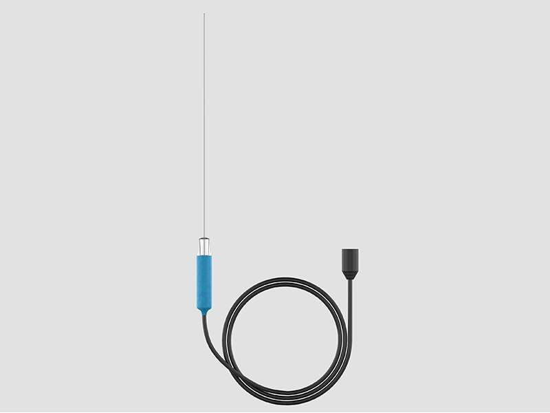
The combination of gauge, length, and exposed tip directly controls your thermal field. These three dimensions dictate how thermal energy disperses through surrounding tissues. If you misjudge these parameters, you severely limit the efficacy of the radiofrequency ablation. A needle too thick might over-extend the burn zone. A tip too short might fail to capture an aberrantly located nerve. Physicians must understand how these physical dimensions intersect to form the desired burn shape. Complete nerve ablation requires placing the densest part of the thermal lesion directly over the target structure.
Patient safety hinges on device integrity. Compromised insulation poses a significant danger during any ablation procedure. If electrical leakage occurs along the needle shaft, you risk causing inadvertent motor nerve injury. Thermal damage to non-target tissue can lead to severe postoperative pain or permanent deficits. Using inappropriate tip lengths similarly endangers surrounding anatomy. A large active tip placed too close to a critical motor pathway might overlap the safe zone. Precise specification matching acts as your first line of defense against collateral tissue damage.
Clinical efficiency improves dramatically when you choose the correct equipment. High-quality devices provide smooth tissue penetration, offering distinct tactile feedback. This feedback helps physicians distinguish between muscle, ligament, and bone. Clear imaging visibility reduces your reliance on multiple needle adjustments. When you can see the needle clearly under ultrasound or fluoroscopy, you reach the target faster. Fewer needle passes directly lower patient trauma. Furthermore, faster targeting significantly reduces radiation exposure for both the patient and the operating room staff.
You must frame your length selection strictly around the depth of the target structures. Using a generic needle size for all patients invites clinical failure. Every target requires a specific reach. Superficial medial branches demand different tools than deep sympathetic chains or dorsal root ganglia. To achieve optimal procedural stability, you must match the RF cannula length to the patient's specific anatomical depth.
Short Cannulae (50mm)
Short needles provide exceptional control for superficial anatomy. They are ideal for cervical medial branch blocks. When working in the delicate cervical spine, precision remains paramount. A 50mm needle minimizes awkward handling. It also strictly limits the risk of advancing too deeply into vulnerable neural or vascular structures. The short shaft keeps the hub close to the skin, preventing unwanted leverage or shifting during the burn.
Medium Cannulae (100mm)
The 100mm needle serves as the workhorse for most pain clinics. It represents the standard size for lumbar medial branch radiofrequency ablation. This length perfectly balances reach and control. It navigates the subcutaneous fat and muscle layers of the average adult back effortlessly. For the vast majority of routine spinal procedures, a medium needle provides exactly the right amount of depth.
Long Cannulae (150mm - 200mm+)
Deep target access demands extended needles. Splanchnic nerves and specific sacroiliac joint approaches require significant reach. You will also need long cannulae when performing radiofrequency ablation on patients with a higher body mass index (BMI). Deep pelvic or abdominal targets require 150mm to 200mm shafts to secure the correct approach angle without depressing the patient's skin excessively.
Needle Length | Primary Clinical Target | Key Advantage |
|---|---|---|
50mm | Cervical medial branches, superficial nerves | Maximum tactile control; prevents over-insertion in delicate areas. |
100mm | Lumbar medial branches, standard spinal targets | Optimal balance of reach and stability for average adult anatomy. |
150mm+ | Splanchnic nerves, deep pelvic targets, high BMI patients | Allows proper trajectory angles for difficult-to-reach internal structures. |
We strongly warn against standardizing on a single length for all procedures. Many facilities attempt to stock only 100mm needles to simplify inventory. This approach compromises patient care. Using excessively long needles for shallow targets causes proximal weight imbalance. The heavy hub extending far outside the skin pulls the needle off target. This imbalance compromises the physician’s tactile feedback. Always match the tool to the specific anatomical requirement.
The active tip defines the very heart of the ablation process. The insulated shaft protects tissue, while the uninsulated portion emits the RF energy. This exposed metal creates a prolate spheroid thermal lesion. The burn zone takes on a football-like shape, radiating outward from the tip. It primarily expands laterally rather than extending straight forward. Understanding this geometry helps you place the needle parallel to the target nerve for maximum capture.
Selecting the RF cannula active tip determines your margin of error. We classify tip exposures into three main categories:
Small Active Tips (2mm - 4mm): These sizes exist for highly localized, precise ablation. You use them when target nerves lie in close proximity to critical motor nerves. The cervical spine often requires these micro-lesions to ensure surrounding motor roots remain completely untouched.
Standard Active Tips (5mm - 10mm): These represent the optimal choice for capturing nerves exhibiting anatomical variability. Physicians commonly use them in lumbar medial branch neurotomy. A 10mm tip creates a broad enough burn zone to guarantee the nerve falls within the ablative field, even if it branches unpredictably.
Large/Specialized Active Tips (15mm+): We use massive tips for large joint denervation. Genicular nerve ablation and complex sacroiliac joint procedures benefit greatly from these sizes. A larger ablation volume successfully denervates wide areas, reducing your need for multiple overlapping lesions.
When assessing different brands, pay close attention to the transition point. This is the exact micro-zone where the active tip meets the insulation. A seamless transition represents high-quality manufacturing. Poorly machined needles leave a small ridge or bump at this junction. This ridge causes tissue drag during insertion. Tissue drag severely distorts the physician's tactile feel and causes unnecessary, painful trauma to the patient. A perfectly smooth transition ensures precise placement and better patient comfort.
Electrical safety heavily depends on dielectric strength. The primary function of the RF cannula coating is to prevent electrical leakage. High dielectric strength keeps the RF energy entirely confined to the active tip. Inferior coatings often look fine to the naked eye but fail under microscopic stress. They can micro-fracture during insertion. These invisible cracks allow energy to escape along the shaft, causing dangerous off-target burns in non-target muscle or skin tissue.
The material covering the shaft changes how the needle behaves. Standard Teflon (PTFE) coatings remain highly cost-effective and provide very smooth insertion. They work well for soft tissue targets. However, advanced procedures require advanced materials. When needles must scrape against bone—such as walking off a facet joint—PTFE can peel or shred. Premium devices use advanced silicone or proprietary medical polymers. These high-grade coatings prove highly durable. They resist peeling even after repeated, aggressive bone contact, ensuring continuous patient safety.
You cannot ablate what you cannot see. Excellent insulation materials often incorporate elements designed specifically for medical imaging.
Ultrasound Guidance: Needles often vanish on ultrasound monitors at steep angles. Look for built-in echogenic enhancements. Premium coatings use specialized micro-etching or specific polymer densities. These features reflect high-frequency sound waves directly back to the probe. This ensures clear, real-time visualization of the entire needle shaft.
Fluoroscopy: X-ray guidance requires distinct contrast. You must ensure both the shaft and the tip provide clear radiopaque outlines. The transition between the insulated shaft and the bare metal tip must show distinctly on the screen. This contrast allows you to verify exact final placement before you initiate the high-heat burn.
Your first procurement step must address capital equipment. Verify that the chosen cannulae connect flawlessly to your existing RF generators. Most modern clinics use machines from Boston Scientific, Stryker, Pajunk, or Nimbus. You must check if the disposable needles feature universal hubs or if they require specific, proprietary connection cables. Buying excellent needles that fail to connect to your generator halts procedures entirely. Standardizing connection types across your facility prevents last-minute clinical delays.
You must connect gauge selection directly to needle rigidity. Thicker gauges, such as 18G, steer remarkably well in dense, fibrotic tissue. They resist bending and create slightly larger lesions due to their larger surface area. However, thicker needles cause more insertion pain. Thinner gauges, like 22G, minimize patient discomfort significantly. They work perfectly for superficial or straightforward approaches. Be aware, though, that 22G needles deflect much more easily when hitting fascial planes. The 20G needle often provides the perfect compromise between steerability and patient comfort.
Procurement must carefully balance unit economics against clinical necessity. Standard disposable needles cost less upfront and work adequately for simple procedures. However, complex cases demand premium features. Specialized echogenic coatings save precious minutes in the operating room. Venom-style tip designs penetrate dense scar tissue easily. You must assess whether the higher unit price of a premium needle offsets the extended operating room time caused by using a cheaper, inferior product. Allocate premium needles to specific, challenging cases to balance the budget effectively.
Do not buy inventory blindly based on paper specifications. We strongly recommend requesting a sample trial for a specific, high-volume procedure. Use the samples for a week of lumbar ablations. Ask your physicians to test the tactile feel during insertion. Inspect the needles after use to check insulation durability upon bone contact. Finally, evaluate the imaging clarity on your specific fluoroscopy or ultrasound screens. Practical trials reveal the true quality of the instrument.
The efficacy of a radiofrequency ablation directly correlates with precision. You must match the cannula’s length to the correct anatomical target depth. You must pair the active tip to the required lesion size to ensure comprehensive nerve capture. Finally, you must select the right coating to guarantee electrical safety and imaging visibility. Ignoring any of these three pillars compromises your clinical outcome.
We encourage thorough clinical evaluation before finalizing any procurement decisions. Bring both your physicians and your purchasing teams to the table. Physicians can validate handling and imaging performance, while procurement can ensure institutional standardization and generator compatibility.
Take action today to optimize your clinical setup. Request a clinical sample, download detailed technical specifications, or consult directly with a device specialist to audit your current inventory. Equipping your team with the right tools ensures safer procedures and more effective pain relief for your patients.
A: It is not recommended. Cervical procedures require shorter cannulae (e.g., 50mm) for precise control and safety, whereas lumbar targets typically require 100mm to accommodate anatomical depth.
A: No. While a larger tip increases the chance of capturing a variable nerve, it also increases the risk of thermal damage to adjacent critical structures. Tip size must be strictly tailored to the specific anatomical safe zone.
A: Standard coatings may blend into surrounding tissue on ultrasound. High-quality cannulae often feature echogenic texturing or specific polymer blends over the shaft and near the tip to reflect ultrasound waves, drastically improving real-time needle tracking.


