
 English
Views: 0 Author: Site Editor Publish Time: 2026-07-22 Origin: Site


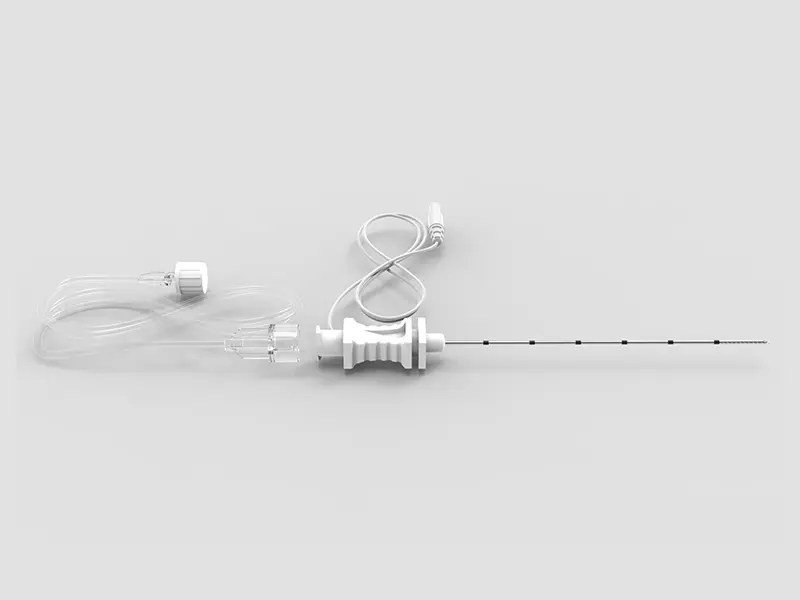
Regional anesthesia has shifted away from landmark-based techniques. Clinicians now rely on precise, ultrasound-guided methods. This major transition fundamentally changes our clinical equipment demands. We no longer rely purely on tactile feedback to find nerve bundles. Visual confirmation is now the standard of care. However, traditional needles lose visibility under ultrasound at steep insertion angles. This invisibility introduces severe clinical risks. Operators might accidentally puncture blood vessels or injure peripheral nerves. You must clearly see your needle tip to ensure patient safety.
This comprehensive guide breaks down the clinical and operational criteria for evaluating echogenic technology. We will help procurement teams and regional anesthesiologists understand the equipment landscape. You will learn how to determine when enhanced-visibility needles become clinically necessary. You will also discover how to balance advanced echogenicity against tactile feedback, injection pressure limits, and per-unit costs. These insights will empower your facility to make confident, evidence-based purchasing decisions.
Echogenic needles utilize specialized surface modifications (like laser etching or corner-cube reflectors) to reflect ultrasound waves back to the probe, significantly improving needle tip and shaft visibility.
The primary clinical driver for adopting an echogenic nerve block needle is maintaining continuous tip visualization during steep-angle insertions or deep target blocks.
While echogenic designs improve safety and procedure efficiency, they do not replace fundamental ultrasound proficiency or tactical feedback requirements.
Evaluating these needles requires balancing advanced echogenicity with tactile "feel," injection pressure limits, and per-unit cost at scale.
Regional anesthesia has transformed over the past two decades. Ultrasound guidance now stands as the definitive gold standard. Previously, practitioners used pure anatomical landmarks. They listened for a physical "pop" through fascial layers. They used nerve stimulators to map anatomical structures. Today, practitioners demand real-time visual confirmation. Seeing the actual nerve structure drastically improves block success rates. It also enhances patient comfort. However, this visual approach introduces a completely new obstacle. We must manage equipment visibility under complex imaging constraints.
During a nerve block needle procedure, you must track the needle tip continuously. Standard metallic needles feature highly polished, smooth surfaces. They act exactly like mirrors under an ultrasound beam. When you insert them at a shallow angle, they reflect sound waves straight back to the transducer. The ultrasound monitor displays a bright, clear image. But clinical reality often requires steep angles. When your insertion angle steepens, an acoustic phenomenon called anisotropy occurs. The smooth surface bounces the ultrasound waves away from the probe. The needle effectively disappears from the monitor screen.
This sudden drop in visibility severely impacts patient safety. Poor visualization drastically increases the probability of clinical errors. Practitioners must never advance a needle blindly. Doing so invites significant risks. We can categorize the most severe risks into three main areas:
Accidental Intraneural Injection: If you cannot see the tip, you might advance it directly into the nerve fascicle. High-pressure injections inside the nerve cause irreversible neurological damage.
Pleural Puncture: Blocks around the neck and chest require extreme precision. Supraclavicular blocks sit very close to the pleura. An invisible tip easily causes a traumatic pneumothorax.
Systemic Toxicity (LAST): Depositing local anesthetics directly into unintended blood vessels triggers local anesthetic systemic toxicity. This condition causes severe cardiovascular and neurological emergencies.
Continuous tip visualization remains your absolute best defense against these complications. Practitioners must maintain a clear field of view from skin puncture to final injection.
Medical facilities must choose the right tools for their specific clinical populations. Comparing various nerve block needle types helps you match clinical requirements to inventory budgets. You generally choose between traditional standard designs and modern echogenic designs. Each type serves a distinct purpose within the operating room.
Manufacturers design standard needles for simple, shallow block applications. They perform beautifully during highly parallel, in-plane approaches. If you keep the needle perfectly perpendicular to the ultrasound beam, standard reflection is highly sufficient. These smooth needles are very cost-effective. They glide effortlessly through soft tissue. However, they rely heavily on the operator's physical skills. You must maintain perfect needle-probe alignment. If your angle steepens or your hand drifts, you lose the ultrasound image entirely.
Engineers build echogenic needles specifically to solve the anisotropy problem. They alter the smooth steel surface. They add microscopic dimples, aggressive grooves, or laser-etched reflectors. These unique patterns catch the descending sound waves. They bounce the acoustic signal directly back to the probe. The needle shines brightly on the monitor regardless of the steep insertion angle. This technology removes the heavy reliance on perfect probe alignment.
Primary use cases for echogenic models typically involve difficult anatomical approaches. Deep blocks, such as sciatic or lumbar plexus blocks, demand highly visible needles. Steep out-of-plane approaches also require them. Additionally, academic training hospitals rely heavily on enhanced echogenicity. Trainees build clinical confidence much faster when they can see their instruments clearly.
Feature | Standard Smooth Needle | Echogenic Enhanced Needle |
|---|---|---|
Surface Texture | Polished, highly smooth metal. | Microscopic dimples, grooves, or laser etching. |
Ultrasound Visibility | Excellent at shallow angles; poor at steep angles. | Consistently bright at all insertion angles. |
Primary Use Case | Superficial blocks, parallel in-plane insertions. | Deep targets, steep angles, out-of-plane approaches. |
Cost Profile | Highly cost-effective per unit. | Premium cost justified by clinical necessity. |
Skill Dependency | Requires strict needle-probe alignment skills. | More forgiving of minor alignment deviations. |
Not all enhanced needles perform equally in clinical settings. Procurement teams and lead physicians must evaluate several specific engineering factors. You must scrutinize these elements before purchasing an echogenic nerve block needle for your facility.
Echogenic Technology and Reflection Quality
First, inspect the actual surface modification method. Some manufacturers use simple laser-etching patterns. Others utilize advanced cornerstone or dimpled reflectors. Cornerstone reflectors trap the sound wave and bounce it back efficiently. You must demand 360-degree echogenicity. Operators frequently rotate the needle hub during insertions. They do this to adjust the bevel direction for optimal anesthetic spread. If the needle only features etching on one side, it will disappear mid-rotation. Full 360-degree coverage ensures the shaft remains visible at all times.
Bevel Design and Tactile Feedback
Ultrasound does not completely replace traditional tactile feedback. Practitioners still rely on the physical "pop" sensation when piercing dense fascial planes. You must evaluate the bevel angle carefully. A sharp 30-degree bevel provides a very distinct pop. This sharp feedback helps you recognize specific tissue layers easily. Conversely, a 15-degree bevel penetrates tissue more smoothly. It causes less overall tissue trauma. However, it offers significantly less tactile response. The right bevel design minimizes nerve trauma while optimizing the spread of the anesthetic fluid.
Fluid Dynamics and Injection Pressure
Needle gauge strictly determines fluid flow limits. A smaller inner diameter restricts flow rates. It drastically increases injection resistance. You must evaluate the inner diameter carefully. Ensure the needle hub supports precise, effortless aspiration. Operators must pull back on the syringe to check for blood before injecting. Integrated extension tubing must also handle sudden pressure changes without ballooning or leaking. Proper fluid dynamics prevent accidental nerve damage caused by high-pressure intrafascicular injections.
Material Rigidity vs. Flexibility
Consider the overall rigidity of the needle shaft. You need the instrument to maintain a perfectly straight trajectory. Passing through dense, fibrous tissue can bend flimsy, low-quality needles. When the shaft bends, it severely misaligns your visual field. The needle tip moves completely out of the two-dimensional ultrasound beam. A sufficiently rigid shaft tracks predictably. It keeps your anatomical target exactly where you expect it to be.
Upgrading your clinical equipment impacts patient outcomes and hospital operations. Administrators need concrete, evidence-based reasons to justify the higher unit price of advanced needles. You must look beyond the initial purchase price. You must focus on the broader operational return on investment.
Clinical Success Criteria: Enhanced visibility directly reduces actual procedure times. Anesthesiologists acquire their anatomical targets much faster. They achieve substantially higher first-pass success rates. They rarely need to withdraw and painfully redirect the needle through the patient's tissue. This improved clinical efficiency keeps demanding operating room schedules running smoothly. Faster blocks mean faster surgical starts. Reduced patient discomfort also leads to higher patient satisfaction scores.
Risk Mitigation & Compliance: Regional anesthesia safety guidelines strongly emphasize continuous tip visualization. National societies publish strict standards regarding nerve block safety. By utilizing an advanced ultrasound guided nerve block needle, you strictly align your facility with these modern best practices. Better visibility actively prevents devastating complications. You spend far less money treating accidental pneumothorax cases. You also avoid highly costly litigation associated with permanent nerve damage or systemic toxicity. Risk mitigation alone justifies the upgrade for complex procedures.
Workflow Scalability: You can effectively standardize these enhanced needles across multiple hospital departments. The Emergency Department, Pain Management clinics, and the primary Anesthesia department can all share the exact same inventory. Standardization significantly simplifies complex ordering processes. It reduces messy storage headaches in supply rooms. More importantly, standardizing high-visibility equipment easily accommodates the widely varying skill levels among your diverse medical staff.
While these advanced tools offer immense clinical benefits, facility-wide adoption presents specific challenges. Clinical directors must manage these risks proactively.
The "False Confidence" Trap: Highly visible equipment does not replace core medical skills. A bright, glowing white line on an ultrasound monitor will not protect the patient if your anatomical knowledge remains poor. Operators still require robust ultrasound fundamentals. They must understand complex patient positioning. They must master transducer ergonomics. They need to interpret the ultrasound anatomy correctly. Do not let advanced equipment breed clinical complacency among your trainees. Continuous education remains vital.
Procurement and Cost Analysis: Echogenic models naturally carry a premium unit cost. Standard alternatives remain significantly cheaper on a per-unit basis. Procurement teams must look past simple line-item pricing. You need to formulate a strong justification model. Base this model on the exact time saved per procedure. Factor in the immense cost of complication avoidance. A single averted permanent nerve injury effectively pays for thousands of upgraded needles. Focus on value, clinical efficiency, and safety outcomes.
Evaluating Vendor Reliability: Always assess your supply chain partners rigorously. You want a consistent, uninterrupted supply of your chosen Nerve Block Needle to prevent sudden workflow disruptions. Ensure the vendor maintains highly reliable stock levels globally. They should offer a comprehensive, wide range of gauges and lengths. Finally, demand excellent vendor-provided clinical training. Your medical staff requires proper onboarding and tactical demonstrations to maximize the distinct benefits of the new equipment.
An echogenic needle easily transitions from a "nice-to-have" luxury item to an absolute clinical necessity. When your regional anesthesia procedures involve steep insertion angles, deep anatomical targets, or highly complex fascial planes, tip visibility becomes non-negotiable. Echogenic technology protects the patient from severe complications. It empowers the clinician to work swiftly and accurately. It ultimately improves the overall efficiency of the operating theater.
Your clinical procurement teams should act decisively. Request a variety of vendor samples immediately. Conduct a tightly controlled trial phase within your facility. Focus this trial exclusively on specific deep blocks, like the lumbar plexus or sciatic nerve. Evaluate the tactile feedback of the bevel alongside the actual ultrasound visibility. Gather structured, honest feedback from your lead anesthesiologists. Use this data to finalize your purchasing decisions before committing to a facility-wide standardization program.
A: Non-echogenic needles feature smooth, polished metal surfaces. They reflect ultrasound waves away from the probe at steep angles, causing poor screen visibility. Echogenic needles use microscopic surface modifications, such as laser-etched grooves or stamped dimples. These unique patterns catch and reflect sound waves directly back to the transducer. This creates a bright, highly visible image regardless of your steep insertion angle.
A: No, clinical guidelines do not universally mandate them for every single procedure. Standard needles work perfectly well for shallow, parallel insertions where visibility remains naturally high. However, guidelines strongly recommend echogenic needles for enhanced patient safety during difficult anatomical approaches. Deep targets and steep out-of-plane insertions strictly require them to maintain continuous tip visualization.
A: Modern echogenic modifications are completely microscopic. They do not significantly alter insertion friction. They also do not increase localized tissue damage compared to standard smooth needles. Awake patients will not feel a difference in physical comfort. The specialized etching strictly alters sound wave reflection without compromising the needle's smooth physical glide through the skin and fascia.
A: Standard sizing typically ranges from 20G to 22G in overall diameter. Lengths usually span from 50mm up to 150mm. You must carefully match the needle size to your specific anatomical target. Shallow, superficial blocks require shorter 50mm lengths. Deep lumbar plexus or transgluteal sciatic nerve blocks demand 100mm to 150mm options to reach the nerve target safely.


